
Surgical Pathology Reporting of Breast Cancer
Dear members of ESP,
Communication of pathology information to the multidisciplinary team caring for cancer patients is essential for good quality clinical management. Careful and accurate surgical pathology reporting of cancer confirms diagnosis, informs about prognosis and helps planning of individualized treatment plans. A well organized and comprehensive report also serves to audit pathology services, improvise service delivery, collect accurate data for cancer registration and epidemiology, facilitate high quality research and provide high quality education. As such, Pathology reports should be accurate, complete, understandable, timely and transferable. The use of standardized formats has been demonstrated to facilitate these requirements and their use has been strongly recommended. They also ensure consistency among institutions and pathologists. In addition, such formats can be supplemented as necessary by the use of free text.
Following the recommendation from the third general assembly of our society, the pathology department in Mekelle University took the assignment of preparing a draft format for surgical pathology reporting of breast cancer. The draft was going to be presented for discussion in the fourth GA which got postponed due to the SARS-Cov-2 pandemic. ESP’s website has an in-built platform to host such discussion where we believe a fruitful dialogue can take place to result in a more refined format for reporting breast cancer.
- These forms include the core components in the reporting of breast cancer and there is no doubt that this will expand with the scope of our practice.
- For the time being, IHC reporting formats are not included here.
- The final synoptic report which will be sent out must include just the dataset question and the response.
- Excisional tissue samples like lumpectomies, wide local excisions or quadrantectomies can also be reported using these formats with modifications.
- Mastectomies after neoadjuvant therapy require special considerations (not included here).
- Institutional variations are not expected to be significant in using these formats.
- CAP, RCPATH and RCPA updated guidelines, in accordance with ICCR, are primarily used to prepare these forms.
Executive council of ESP recognizes and appreciates the staff (Dr. Sara Kiros, Dr. Hadas W/zgina, Dr. Melisachew Mulatu & Dr. Samrawit G/Medhin) of the department of pathology in Mekele University for preparing this draft.
On a final note, I’d like to emphasize that the comments and suggestions of ESP members are critical to the development of a standard format that can be harmonized for national use for better management of patients with breast cancer. It will also maximize the quality of data compilation for epidemiology, research and education.
We will subsequently have similar discussion for reporting of Cervical Cancer and Colorectal cancer.
Meron Demelash, MD
CME/CPD/Publications Coordinator of ESP
Breast Histopathology Request Form
Patient information
Name: ______________________________________________ Gender: ______ Age: ______MRN:_____________
Address:_______________________________________Family Hx of Cancer: __________________________________
Clinical Information(relevant Hx, physical, lab and Imaging findings): ___________________________________
______________________________________________________________________________________
Pre-Op Pathology Result (diagnosis and number)
- Cytology(FNAC):______________________________________________
- Histopathology(needle-core, incisional or excisional):______________________________________________
Specimen Information
o Laterality(Rt or Lt or Bilateral): _________________
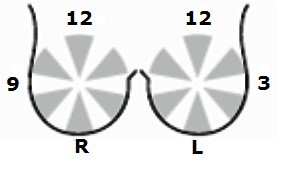
o Position(by quadrant or O’clock): _______________
o Tumor size (by P/E and/or Imaging): _____________
o Procedure: Incision/Excision (WLE, Lumpectomy)/Modified Mastectomy/Radical Mastectomy/ Partial Mastectomy/completion Mastectomy
o Margin Marking done?(Yes or No): __________
o Marking by (sutures, clips etc): __________________________
o Axillary Dissection done? (Yes or No): ________
Presumptive Clinical Diagnosis:_____________________________________________________________________
Preoperative Clinical Stage: ___________ Neoadjuvant Treatment? (Yes or No):______________
New Primary Cancer or Recurrence?:________________
Receptor (ER, PR, HER2) study required?(Yes or No):________________
Physician’s Name and Signature:_____________________________________ Cell phone No: __________________
Date:___________________
Note:
- Fill request form properly to facilitate histologic interpretation
- Put tissue in ample amount (at least twice the volume of tissue) 10 % buffered formalin
- Do not transect tissue as this will interfere with critical components of interpretation like margin assessment
- If you need assistance, feel free to call the pathology laboratory
Grossing Template for Primary Mastectomy Specimen _ IBC
Right/Left Modified Mastectomyspecimen is receivedin formalin in a container labeled with the patient’s name, age and MRN (___________). Specimen measures____ × ____ × ___cm without/with axillary tail of ____ × ____ × ___cm and weighs ____grams. Ellipse of skin and nipple is ____ × ____ cm. The skin is grossly unremarkable/or is remarkable for ____________________. The nipple and areola complex are grossly unremarkable/or is remarkable for _______________.The breast is serially sectioned sequentiallyinto____ slices with the nipple located in slice number ____.The cut surface reveals one/multiple (Specify:_____) circumscribed/irregular tan white tumor ____________________________________
on slice numbers____, ____, and ____ in the ________ quadrant (largest) measuring ____× ____ × ____cm. The lesion is ____cm from the superior margin, _____cm from the inferior margin,_____cm from the medial margin _____cm from the lateral margin, ____cm from the deep margin, and____cm from the skin. The remainder of the parenchyma is fibro-fatty that is unremarkable/remarkable for __________________________. The total number of non-sentinel axillary LNs harvested is_______with size ranging from ___ × ___ cm to ___ × ___cm.Matted LNs are present/absent. Skeletal muscleis/is not present. Other smaller tumor (in multifocal lesions) or relevant gross pathological findings include________________________________________________________
Sample Block Key
A-D: 3-4 representative areas of the tumor with rim of peritumoral tissue
E-F: Margins (include all margins less than 10mm from tumor)
G-I: Intervening tissues in multifocal lesions
J: Non-tumor lesion (if recognizable)
K-N: Non-lesional tissue from each quadrant
O-P: Nipple and abnormal skin, if present
Q+: Lymph nodes
SP Number: …………………..Histopathological Reporting of Invasive Breast Cancer in Surgical Resections
Patient Name: …………………………..………………………..…….… Age:…….… Sex: …….…. MRN:………………….. Sending Institutions: ……………………………………Date of Surgery: ………………… Date of Reporting: ………………
Laterality: Right □ Left □
Specimen type (Procedure): Incisional Biopsy □ Excision Biopsy □ Modified Mastectomy □ Radical Mastectomy □ Partial Mastectomy □ Completion Mastectomy □
Largest invasive tumour Size (mm): ............
Focality: Unifocal □ Multifocal □ Cannot be assessed □
Histologic Type: Pure □ (select one box below) Mixed □ (select all components present below)
Ductal/NST □ Tubular/cribriform □ Lobular □ Mucinous □ Medullary-like □ Micropapillary □
Othertype/component: .................................................................................................................................
Histological grade 1 □ 2 □ 3 □ Cannot be assessed □.......................................................
Lymphovascular invasion Present □ Not identified □ Uncertain □Cannot be assessed □
Lymph node Status :
Axillary nodes: Total present: .......... Total positive: ...........
For single node positive: Macrometastasis □ Micrometastasis □ ITCs □
Other nodes: Site: .............................Total present: .......... Total positive:. ..........
For single node positive: Macrometastasis □ Micrometastasis □ ITCs □
In situ component(s)
DCIS: Present □ Not identified □ Cannot be assessed □
DCIS grade: High □ Intermediate □ Low □ Cannot be assessed □
Tumor extension (skin, nipple and/or skeletal muscle): …………………………………….……………………………………..
TNM stage (AJCC 8th Edition)
T stage: pTis □ pT1mi □ pT1a □ pT1b □ pT1c □ pT2 □ pT3 □ pT4a □ pT4b □
pT4c □ pT4d □ Cannot be assessed □
N stage: pN0 □ pN1mi □ pN1a □ pN1b □ pN1c □ pN2a □ pN2b □ pN3a □
pN3c □ Cannot be assessed □
M stage: pM0 pM1 □ Cannot be assessed □
Surgical Margin Status Positive □ Negative □
Which margin is positive? Superior □ Inferior □ Medial □ Lateral .□
Deep□ Superficial □ Nipple margin□
Other pathological findings: ………………………………………………..……………………………………………………………………
Summary Diagnosis
Laterality/Procedure: Grade, Histologic Type, TNM Stage, Margin Status
COMMENT
When result of a receptor status study is available, a second report including the result as an addendum will be issued.
Sample Final Report
SP Number: 1234/20
Histopathological Report
Patient Name: De-identified De-identified Age:58 Sex: F MRN:123456 Sending Institutions: Ayder Comprehensive Specialized Hospital
Date of Surgery: 13/08/20 Date of Reporting: 25/08/20
Laterality: Right
Specimen/procedure type: Modified Mastectomywith Axillary Dissection
Largest invasive tumour Size (mm): 65(greatest dimension)
Focality:Unifocal
Histologic Type:Pure, Invasive Carcinoma, NST
Histological grade: 3 (poorly differentiated)
- Tubule formation=2, Nuclear grade=3, and Mitoses=3
Lymphovascular invasion:Present
Lymph node Status :
Axillary nodes: Total present: 12 Total positive: 4
Type of LN metastasis: Macrometastasis
Other nodes: Not received
DCIS:Present
DCIS grade:High, solid with necrosis
Tumor extension (skin, nipple and/or skeletal muscle): Absent
TNM stage (AJCC 8th Edition)
T stage: pT3
N stage: pN2a
M stage: Cannot be assessed
Surgical Margin Status: Positive, DeepMargin
Other pathological findings: Fibrocystic change
Summary Diagnosis
Breast, Right Modified Mastectomy: Poorly Differentiated Invasive Carcinoma (NST), pT3N2aMx(Positive deep margin)
COMMENT
When result of a receptor status study is available, a second report including the result as an addendum will be issued.